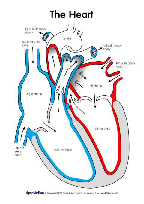
40 structure of the heart without labels
Parts and Components of Human Ear and Their Functions In addition to helping the body take in auditory messages, the ear helps to maintain a proper head position. The fluid in the ear also helps the body maintain a sense of balance so the body can maintain proper posture and coordination. There are three major parts of the ear, the outer, middle and inner ear. Each contains several parts that are ... Parts of the Body - Vocabulary heart - your heart pumps your blood around your body. lungs - when you breathe, the air goes into your lungs. veins - these transport blood through your body. They are like little tubes. brain - this is your 'thinking machine' inside your head. throat - food goes down this to get to your stomach. liver - the organ that cleans your blood.
The power of ambition | Lead Stories | Jamaica Gleaner Fifty-three-year-old Dezreen Miller embraces her 23-year-old daughter, Anita Stephenson, in front of their home in Olympic Gardens, St Andrew. After living in the partially collapsed one-room structure for years, they are hopeful that better days are on the horizon as Stephenson is set to start her first job as a teacher come next month ...
Structure of the heart without labels
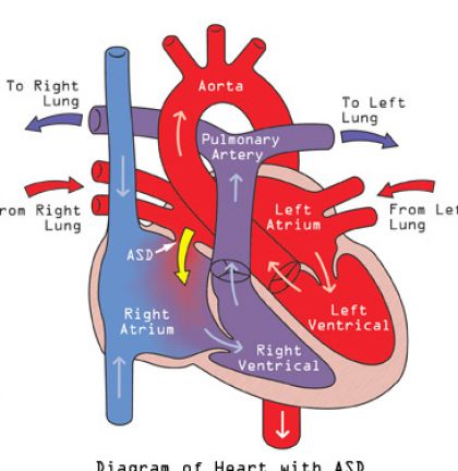
› heart-healthHeart Health | Heart Attack Prevention | Bayer® Aspirin TO HELP PREVENT ANOTHER HEART ATTACK. A doctor-directed aspirin regimen helps keep your blood flowing. Along with other heart-healthy choices, it can reduce your risk of having another heart attack. Learn About Aspirin's Benefits. Aspirin is not appropriate for everyone, so be sure to talk to your doctor before you begin an aspirin regimen. hbr.org › 2009 › 09How Strategy Shapes Structure - Harvard Business Review Summary. Reprint: R0909H. When executives develop corporate strategy, they nearly always begin by analyzing the industry or environmental conditions in which they operate and the strengths and ... Diagram of Human Heart and Blood Circulation in It The wall of the heart has three different layers, such as the Myocardium, the Epicardium, and the Endocardium. Here's more about these three layers. Epicardium. The outermost layer of your heart wall is called the epicardium, which is basically a very thin layer of serous membrane.
Structure of the heart without labels. They Named Me The 'Most Notorious Card Counter in America.' This Is The ... A casual conversation led to a meeting which led to joining a team of card-counting parishioners, and together we beat casinos at the game of blackjack from coast to coast for seven years, banked by investors for a million dollars. Casinos would eventually label me the "most notorious card counter in America." Capillaries - Structure & Function Explained with Diagrams - TeachPE.com Structure of Capillaries. Capillaries have very thin walls comprised only of endothelial cells, which allows substances to move through the wall with ease. Capillaries are very small, measuring 5-10 micrometres in width. However, the cross-sectional area of capillaries within an average size muscle would be larger than that of the Aorta. Aortic valve stenosis - Diagnosis and treatment - Mayo Clinic It can help determine whether the heart is enlarged, which can occur in aortic valve stenosis. It can also show swelling of the aorta and calcium buildup on the aortic valve. Exercise tests or stress tests. These tests often involve walking on a treadmill or riding a stationary bike while the heart is monitored. Corpus callosum: Anatomy, function and clinical aspects | Kenhub The corpus callosum is a large white matter tract that connects the two hemispheres of the brain. It is an incredibly important structural and functional part of the brain. It allows us to perceive depth and enables the two sides of our brain to communicate. The corpus callosum gets its name from the Latin language ("tough body").
Arthropod - Wikipedia The heart is typically a muscular tube that runs just under the back and for most of the length of the hemocoel. It contracts in ripples that run from rear to front, pushing blood forwards. Sections not being squeezed by the heart muscle are expanded either by elastic ligaments or by small muscles, in either case connecting the heart to the body wall. Along the heart run a series of paired ostia, non-return valves that allow blood to enter the heart but prevent it from leaving before it ... Antenatal Care Module: 6. Anatomy of the Female Pelvis and Fetal Skull ... 6.2.1 The size and shape of the pelvis. The size and shape of the pelvis is important for labour and delivery. Well-built healthy women, who had a good diet during their childhood growth period, usually have a broad pelvis that is well adapted for childbirth. It has a round pelvic brim and short, blunt ischial spines. Stereotactic Body Radiotherapy (SBRT): Uses, Side Effects, Procedure ... Adjacent organs: SBRT is also typically avoided if it can cause harm to any organ or structure critical to the body's function. This includes the heart, major vessels, spinal cord, brachial plexus, phrenic nerve, and recurrent laryngeal nerve. SBRT should only be considered if these structures are at least 2 cm away from the tumor. › design-templates › printHeart Diagram – 15+ Free Printable Word, Excel, EPS, PSD ... For every use a template has been designed with a motive of making it easy for the user to get the print of it without making a new one of his own. You may also visit venn diagram templates. Teachers and students use the heart diagram, in biological science, to study the structure and functions of a human being’s heart.
Atom - Wikipedia An atom is the smallest unit of ordinary matter that forms a chemical element. Every solid, liquid, gas, and plasma is composed of neutral or ionized atoms. Atoms are extremely small, typically around 100 picometers across. They are so small that accurately predicting their behavior using classical physics, as if they were tennis balls for example, is not possible due to quantum effects. My adult ADHD drugs felt like a lifeline. Then came the scary side ... Without the structure of regular mealtimes and bedtimes, my rhythms became erratic. ... clothes labels no longer bothered me to distraction. ... It also made my heart race - once, I had a heart ... Circulatory System Diagram | New Health Advisor Heart consists of four chambers namely right atria, left atria, right ventricle and left ventricle. Both the atrium and ventricles are separated from each other with a muscular septum. Valves are present in between atria and ventricle which helps in draining the blood from upper part to lower part of the body. The 9/11 Memorials and Artifacts Throughout NYC 10. Union Square Subway Station Memorial. On the white subway subway station and pasted the transparent mailing labels on the tiled wall. The MTA left the memorial in place, although after so many ...
Label the heart - Teaching resources
Researchers boost sensitivity and speed of Raman microscopy technique ... Researchers have developed a label-free and non-invasive Raman spectroscopy approach that can acquire microscopic images of biological samples and identify a wide range of biomolecules with ...
Label the heart - Teaching resources
heart | Structure, Function, Diagram, Anatomy, & Facts The heart cavity is divided down the middle into a right and a left heart, which in turn are subdivided into two chambers. The upper chamber is called an atrium (or auricle), and the lower chamber is called a ventricle. The two atria act as receiving chambers for blood entering the heart; the more muscular ventricles pump the blood out of the heart.

Labeled picture of the heart – Graph Diagram
Allen Balik, The Wine Exchange: Complexity beyond flavor and aroma A wine's structure is an extension of balance as they both rely on similar components expressing themselves in a slightly different manner. Acidity forms the backbone (or skeleton) of the wine ...
Picture of the heart labeled
Step Inside the Delhi Home of an Indian Princess August 20, 2022. Ashish Sahi. In a leafy 1950s neighborhood of garden bungalows set in symmetrical squares adjoining the Delhi zoo, the home of Priti Pratap Singh, Princess of the erstwhile ...
Related Items
Organelle - Genome.gov Definition. …. An organelle is a subcellular structure that has one or more specific jobs to perform in the cell, much like an organ does in the body. Among the more important cell organelles are the nuclei, which store genetic information; mitochondria, which produce chemical energy; and ribosomes, which assemble proteins.
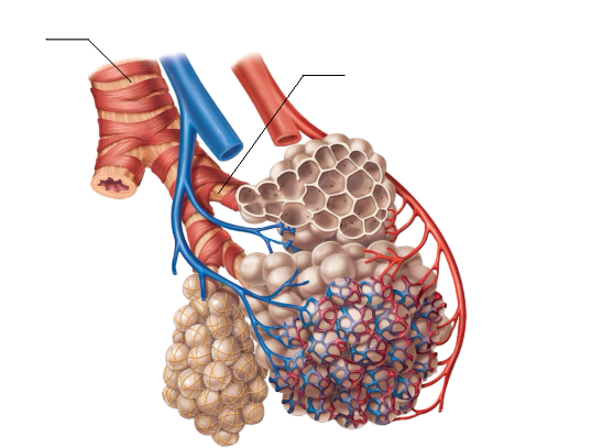
Respiratory system jnr
Diaphragm: Function, Anatomy, and Abnormalities - Verywell Health Anatomy. The diaphragm is a parachute-shaped fibrous muscle that runs between the chest and abdomen, separating these two large cavities. It is asymmetric, as its right dome is larger than the left dome. The diaphragm has openings that allow certain structures to span the chest and abdominal cavities.
Post a Comment for "40 structure of the heart without labels"